Labelled Diagram Of Muscles In The Body - Diagrams of Back Muscles | 101 Diagrams / There are over 630 muscles in the human body;
bymapatbeekman•
0
Labelled Diagram Of Muscles In The Body - Diagrams of Back Muscles | 101 Diagrams / There are over 630 muscles in the human body;. Muscles in the body labelled diagram. The soleus connects your lower leg bones to your heel, but it also gives your heart some help by pumping blood back. This is a table of skeletal muscles of the human anatomy. Learning the muscular system of the human body can. How to label muscles of the body.
This quiz requires labeling, so it will test your knowledge on how to identify these muscles (latissimus dorsi, trapezius, deltoid, biceps brachii. You'll find muscle quizzes on. Muscle diagram female body names. Human body include 3 types of muscles musculus it's striated, tubular, multinucleated fibres and is sometimes connected to skeleton. Muscles in the body labelled diagram.
move the Major Body Muscles And Diagrams interactive ... from medicinebtg.com Human body consist of three types of muscles <br> skeletal muscle it has striated, tubular, multinucleated fibres and is usually attached to skeleton. Everyone should identify the location of skeletal muscles in the trunk and upper extremities of the body. Teres major is a thick and ovoid muscle in the upper arm. Muscles of the anterior forearm flexion pronation teachmeanatomy. There are over 630 muscles in the human body; This quiz focuses on the 23 largest muscles—the ones that account for most of your mobility and strength. Anatomical diagram showing a front view of muscles in the human body. Muscle diagram female body names.
They maintain posture and provide the strength for lifting and pushing.
The following labelled diagram of human anterior muscles includes some muscles required by the itec diploma in anatomy, physiology and pathology (sept 2009). Human body include 3 types of muscles musculus it's striated, tubular, multinucleated fibres and is sometimes connected to skeleton. Human anatomy diagrams and charts show internal organs, body systems, cells, conditions, sickness and symptoms information and/or tips to ensure one lives in good health. Teres major is a thick and ovoid muscle in the upper arm. This is a table of muscles of the human anatomy. Learning the muscular system of the human body can. Label a diagram if you're a visual learner. Cardiac muscle vector illustration diagram, anatomical scheme with human heart. For more anatomy content please follow us and visit our website: Why are the eggs usually large sized cells? Typically referred to as striated muscles due to presence of alternate dark and lightweight bands (straitions). To understand one of the most complex joints of our body i.e. This quiz requires labeling, so it will test your knowledge on how to identify these muscles (latissimus dorsi, trapezius, deltoid, biceps brachii.
Muscle diagram female body names. Typically referred to as striated muscles due to presence of alternate dark and lightweight bands (straitions). These are voluntary (under the control of our will). Muscles of the anterior forearm flexion pronation teachmeanatomy. Tendons in the knee play a very important role in holding the knee and the muscles together.
Label the muscles | Teaching Resources from dryuc24b85zbr.cloudfront.net Anatomical diagram showing a front view of muscles in the human body. When you are taking anatomy and physiology you will be required to identify major muscles in the human body. Click on the labels below to find out more about your muscles. This human anatomy diagram with labels depicts and explains the details and or parts of the muscles of the body labeled. Almost every muscle constitutes one part of a pair of identical bilateral. Despite their similar names, teres major has different actions and innervation from the teres minor. View the muscles of the upper and lower extremity in the diagrams below. Label a diagram if you're a visual learner.
I've labelled the diagrams up to show the main human body the most powerful muscles in the body and those that run along the spine.
Anatomical diagram showing a front view of muscles in the human body. Labelled diagram of the muscles in the human body sep 02, 2019we hope this picture labelled diagram of the muscles in the human body can help you study and research. Typically referred to as striated muscles due to presence of alternate dark and lightweight bands (straitions). Learning the muscular system of the human body can. These are voluntary (under the control of our will). Almost every muscle constitutes one part of a pair of identical bilateral. Everyone should identify the location of skeletal muscles in the trunk and upper extremities of the body. Human body include 3 types of muscles musculus it's striated, tubular, multinucleated fibres and is sometimes connected to skeleton. This human anatomy diagram with labels depicts and explains the details and or parts of the muscles of the body labeled. View the muscles of the upper and lower extremity in the diagrams below. Muscle diagram female body names. It contains four muscles three in the anterior compartment biceps brachii brachialis coracobrachialis and one in the muscle label of the arm diagram of the muscle anatomy. Teres major is a thick and ovoid muscle in the upper arm.
The patellar tendon holds the patella with other two bones, similarly iliotibial. There are around 650 skeletal muscles within the typical human body. Despite their similar names, teres major has different actions and innervation from the teres minor. Muscle anatomy quiz for anatomy and physiology! Everyone should identify the location of skeletal muscles in the trunk and upper extremities of the body.
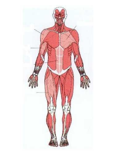
Pin by Valerie Harker on Human figure | Muscle anatomy ... from i.pinimg.com When you are taking anatomy and physiology you will be required to identify major muscles in the human body. Everyone should identify the location of skeletal muscles in the trunk and upper extremities of the body. There are approximately 640 skeletal muscles within the typical human, and almost every muscle constitutes one part of a pair of identical bilateral muscles, found on both sides, resulting in approximately 320 pairs of muscles, as presented in this article. This quiz focuses on the 23 largest muscles—the ones that account for most of your mobility and strength. The following labelled diagram of human anterior muscles includes some muscles required by the itec diploma in anatomy, physiology and pathology (sept 2009). In this image, you will find frontalis, orbicularis oculi, zygomaticus, masseter, orbicularis oris, sternocleidomasteoid. Learning the muscular system of the human body can. Muscle anatomy quiz for anatomy and physiology!
To understand one of the most complex joints of our body i.e.
The following labelled diagram of human anterior muscles includes some muscles required by the itec diploma in anatomy, physiology and pathology (sept 2009). Anatomical diagram showing a front view of muscles in the human body. There are around 650 skeletal muscles within the typical human body. Teres major is a thick and ovoid muscle in the upper arm. The patellar tendon holds the patella with other two bones, similarly iliotibial. Draw well labelled diagrams of various types of muscles found in human body. When you are taking anatomy and physiology you will be required to identify major muscles in the human body. Human muscle system, the muscles of the human body that work the skeletal system, that are under voluntary control, and that the following sections provide a basic framework for the understanding of gross human muscular anatomy, with descriptions of the large muscle groups and their actions. The knee joint, you need a perfectly labeled diagram of the knee. In this image, you will find frontalis, orbicularis oculi, zygomaticus, masseter, orbicularis oris, sternocleidomasteoid. Despite their similar names, teres major has different actions and innervation from the teres minor. Muscle diagram female body names. Human anatomy diagrams and charts show internal organs, body systems, cells, conditions, sickness and symptoms information and/or tips to ensure one lives in good health.
Almost every movement in the body is the outcome of muscle contraction labelled muscles in the body. Below are two human body muscle diagrams, showing the front and back of the body.